Bursitis Of The Feet Treatment
Overview
Bursae are situated in various locations throughout the body where friction between tissues commonly occurs. These sacs are designed to help reduce friction and prevent pain. Repetitive movements or prolonged and excessive pressure are the most common causes of bursal inflammation, though traumatic injury may also cause this painful problem. In fact, the body sometimes creates bursal sacs in response to trauma or tissue damage.
Causes
There are several factors which can predispose patients to developing this condition. These need to be assessed and corrected with direction from a physiotherapist and may include poor foot biomechanics (particularly flat feet), inappropriate footwear (e.g. excessively tight fitting shoes), muscle weakness (particularly the calf, quadriceps and gluteals), muscle tightness (particularly the calf), joint stiffness (particularly the ankle, subtalar joint or foot), bony anomalies of the heel bone, inappropriate or excessive training or activity, inadequate recovery periods from sport or activity, inadequate warm up, inadequate rehabilitation following a previous Achilles injury, change in training conditions or surfaces, inappropriate running technique, inadequate fitness, poor pelvic and core stability, poor proprioception or balance, being overweight.
Symptoms
Where the tendon joins the calcaneal bone, friction can cause the spaces between the tendon, bone and skin to swell and inflame with bursitis. This constitutes a calcaneal bursa. Apart from swelling over the back of the heel, you?ll feel acute tenderness and pain when you move it or even apply light pressure. Your swollen heel may look more red than the other one, and the swelling is often so hard it can feel like bone, partly because it sometimes is, as a bony overgrowth can occur in chronic cases.
Diagnosis
During the physical examination of a patient with calcaneal bursitis, the physician should keep the following considerations in mind. Swelling and redness of the posterior heel (the pump bump) may be clearly apparent. The inflamed area, which may be slightly warm to the touch, is generally tender to palpation. Careful examination can help the clinician to distinguish whether the inflammation is posterior to the Achilles tendon (within the subcutaneous calcaneal bursa) or anterior to the tendon (within the subtendinous calcaneal bursa). Differentiating Achilles tendinitis/tendinosis from bursitis may be impossible. At times, the 2 conditions co-exist. Isolated subtendinous calcaneal bursitis is characterized by tenderness that is best isolated by palpating just anterior to the medial and lateral edges of the distal Achilles tendon. Conversely, insertional Achilles tendinitis is notable for tenderness that is located slightly more distally, where the Achilles tendon inserts on the posterior calcaneus. A patient with plantar fasciitis has tenderness along the posterior aspect of the sole, but he/she should not have tenderness with palpation of the posterior heel or ankle. A patient with a complete avulsion or rupture of the Achilles tendon demonstrates a palpable defect in the tendon, weakness in plantarflexion, and a positive Thompson test on physical examination. During the Thompson test, the examiner squeezes the calf. The test is negative if this maneuver results in passive plantarflexion of the ankle, which would indicate that the Achilles tendon is at least partially intact.
Non Surgical Treatment
With posterior Achilles tendon bursitis, treatment is aimed at reducing the inflammation and adjusting the foot's position in the shoe to relieve pressure and motion on the back of the heel. Foam rubber or felt heel pads can be placed in the shoe to eliminate pressure by elevating the heel. Placing protective gel padding over the painful bursa or stretching the back part of the shoe and placing padding around the inflamed bursa may help. Sometimes a special shoe, such as a running shoe designed to stabilize the midsole heel, devices placed in the shoe (orthoses), or both can help to control abnormal foot and heel motion contributing to the posterior heel irritation. Other shoes have padding that reduces irritation to the posterior heel and Achilles tendon.
Surgical Treatment
Surgery is rarely need to treat most of these conditions. A patient with a soft tissue rheumatic syndrome may need surgery, however, if problems persist and other treatment methods do not help symptoms.
Prevention
You can avoid the situation all together if you stop activity as soon as you see, and feel, the signs. Many runners attempt to push through pain, but ignoring symptoms only leads to more problems. It?s better to take some time off right away than to end up taking far more time off later. Runners aren?t the only ones at risk. The condition can happen to any type of athlete of any age. For all you women out there who love to wear high-heels-you?re at a greater risk as well. Plus, anyone whose shoes are too tight can end up with calcaneal bursitis, so make sure your footwear fits. If the outside of your heel and ankle hurts, calcaneal bursitis could be to blame. Get it checked out.
Hammertoe Surgery Treatment
 Overview
A Hammer toe can be flexible or rigid. Hammertoes often start out flexible and become rigid over time as your toe becomes accustomed to its crooked position. Flexible hammertoes are less serious than rigid hammertoes, as they can be easily diagnosed and treated in their initial stages. Flexible hammertoes are named as such because your affected toe still possesses some degree of movement.
Causes
Some causes of hammertoe are shoes that are too tight or short, shoes with high heels, injury, Diseases that affect the nerves and muscles, such as arthritis and diabetes. When shoes do not fit well, over time the pressure of the shoes pushes the toes into a bent position. After a while, the muscles become unable to straighten the toe, even when you are not wearing shoes. Similarly, when there is damage or disease of the nerves or muscles in the toes, the toe may rest in the bent position until the tendons become permanently shortened and the toe becomes a rigid hammertoe. The risk of developing a hammertoe increases with age. Women are much more likely to develop a hammertoe than men.
 Symptoms
A soft corn, or heloma molle, may exist in the web space between toes. This is more commonly caused by an exostosis, which is basically an extra growth of bone possibly due to your foot structure. As this outgrowth of excessive bone rubs against other toes, there is friction between the toes and a corn forms for your protection.
Diagnosis
Your healthcare provider will examine your foot, checking for redness, swelling, corns, and calluses. Your provider will also measure the flexibility of your toes and test how much feeling you have in your toes. You may have blood tests to check for arthritis, diabetes, and infection.
Non Surgical Treatment
Changing the type of footwear worn is a very important step in the treatment of hammer toes. When choosing a shoe, make sure the toe box (toe area) is high and broad, and can accommodate the hammer toes. A shoe with a high, broad toe box will provide enough room in the forefoot area so that there is less friction against the toes. Other conservative treatments include using forefoot products designed to relieve hammer toes, such as hammer toe crests and hammer toe hammertoe splints. These devices will help hold down the hammer toe and provide relief to the forefoot. Gel toe shields and gel toe caps are also recommended to eliminate friction between the shoe and the toe, while providing comfort and lubrication.
Surgical Treatment
Curative treatment of hammertoes varies depending upon the severity of the deformity. When the hammertoe is flexible, a simple tendon release in the toe works well. The recovery is rapid often requiring nothing more that a single stitch and a Band-Aid. Of course if several toes are done at the same time, the recovery make take a bit longer.
 Prevention
Prevention of a hammer toe can be difficult as symptoms do not usually start until the problem is well established. Wearing shoes that have extra room in the toes may help the problem or slow down its development.
Why Do I Have Bunions?
Overview
 Hallux valgus is a condition that affects the joint at the base of the big toe. The condition is commonly called a bunion. The bunion actually refers to the bump that grows on the side of the first metatarsophalangeal (MTP) joint. In reality, the condition is much more complex than a simple bump on the side of the toe. Interestingly, this condition almost never occurs in cultures that do not wear shoes. Pointed shoes, such as high heels and cowboy boots, can contribute to the development of hallux valgus. Wide shoes, with plenty of room for the toes, lessen the chances of developing the deformity and help reduce the irritation on the bunion if you already have one.
Causes
Various factors, including a tight gastrocnemius (or calf) muscle and instability of the arch, contribute to formation of bunions. The tight calf muscle is often hereditary and can cause a bunion because it forces more loading, or pressure, on the forefoot. Ultimately, this can contribute to instability in the bones, ligaments and tendons that form the arch. When it?s unstable, the arch starts collapsing and the metatarsal can shift. Arch instability can also be brought on by obesity, again, due to chronic overloading of the foot. But, by far, the most common contributing factor is childbirth. Bunions are most common in women who have had children. This happens because the hormones that affect their pelvis during childbirth also affect their feet. The hormone is called relaxin, and it allows bones to move and spread. Over time, it can cause the structure of a woman?s feet to gradually stretch and the metatarsal to shift.
Symptoms
Redness, swelling, or pain along the inside margin of the foot just behind the great toe. Moderate to severe discomfort at the bunion when wearing shoes, particularly if tight fitting. A painful callus may develop over the bunion. Sometimes a painful corn on the adjacent sides of the first and second toes. Irritation if there is overlapping of the first and second toes. Arthritis may cause stiffness and discomfort in the joint between the great toe and the first metatarsal. There may be a fluid filled cyst or bursa between the skin and the "bunion bone". Skin over the bunion may break down causing an ulceration, which can become infected.
Diagnosis
Physical examination typically reveals a prominence on the inside (medial) aspect of the forefoot. This represents the bony prominence associated with the great toe joint ( the medial aspect of the first metatarsal head). The great toe is deviated to the outside (laterally) and often rotated slightly. This produces uncovering of the joint at the base of the big toe (first metatarsophalangeal joint subluxation). In mild and moderate bunions, this joint may be repositioned back to a neutral position (reduced) on physical examination. With increased deformity or arthritic changes in the first MTP joint, this joint cannot be fully reduced. Patients may also have a callus at the base of their second toe under their second metatarsal head in the sole of the forefoot. Bunions are often associated with a long second toe.
Non Surgical Treatment
Your podiatrist can recommend exercises, orthoses (special devices inserted into shoes), shoe alterations or night splints (which hold toes straight over night) which may slow the progression of bunions in children. According to experts, ?conservative? measures such as these may help relieve symptoms, though there is no evidence they can correct the underlying deformity. Orthoses are designed to prevent the problem getting worse by decreasing any biomechanical causes of bunions. In other words, if the biomechanical theory is correct (i.e. if your bunions are caused by the way you walk), orthoses may help you walk in a way that doesn?t exacerbate the problem. But it won?t change the already established shape of your foot. For that, you need surgery. 
Surgical Treatment
For severe bunions, outpatient surgery may be recommended. Within hours after surgery, you?ll be on your way home and ready for recovery. Your foot will be bandaged following surgery and placed in a surgical shoe which allows you to remain mobile. Immediate weight bearing without the use of casting or crutches is standard post- operative recovery for bunions. In most cases, the majority of healing should occur within a few weeks and you can resume normal activity within a short period of time. Bunion surgery can both reduce pain and improve the appearance of your feet. After surgery it is important to see your podiatrist as scheduled and follow all recovery instructions.
Prevention
The best way to reduce your chances of developing bunions is to wear shoes that fit properly. Shoes that are too tight or have high heels can force your toes together. Bunions are rare in populations that don?t wear shoes. Make sure your shoes are the correct size and that there's enough room to move your toes freely. It's best to avoid wearing shoes with high heels or pointed toes.
How Do I Know If I Have Over-Pronation Of The Feet
Overview
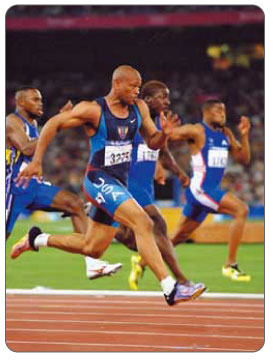
If you've been running long enough, you've most likely had to deal with various aches and pains in your feet, knees, hips or lower back. Plantar fasciitis, achilles tendinitis, IT-band syndrome, meniscus tears, runner's knee, bursitis of the hip or knee, patellofermoral pain syndrome, chondromalacia patella, lower back pain and piriformis syndrome are only some of many conditions an athlete may develop during the course of his or her running career.
Causes
During our development, the muscles, ligaments, and other soft tissue structures that hold our bones together at the joints become looser than normal. When the bones are not held tightly in place, the joints are not aligned properly, and the foot gradually turns outward at the ankle, causing the inner ankle bone to appear more prominent. The foot moves in this direction because it is the path of least resistance. It is more difficult for the foot to move in the opposite direction (this is called supination). As we develop, the muscles and ligaments accommodate to this abnormal alignment. By the time growth is complete, the pronated foot is: abnormally flexible, flat, and its outer border appears raised so that as you step down you do not come down equally across the entire foot; instead, you come down mostly on the inner border of the foot. Normal aging will produce further laxity of our muscles that causes the pronation to become gradually worse.
Symptoms
When standing, your heels lean inward. When standing, one or both of your knee caps turn inward. Conditions such as a flat feet or bunions may occur. You develop knee pain when you are active or involved in athletics. The knee pain slowly goes away when you rest. You abnormally wear out the soles and heels of your shoes very quickly.
Diagnosis
People who overpronate have flat feet or collapsed arches. You can tell whether you overpronate by wetting your feet and standing on a dry, flat surface. If your footprint looks complete, you probably overpronate. Another way to determine whether you have this condition is to simply look at your feet when you stand. If there is no arch on the innermost part of your sole, and it touches the floor, you likely overpronate. The only way to truly know for sure, however, is to be properly diagnosed by a foot and ankle specialist.
Non Surgical Treatment
Studies have shown that the most effective way to dexrease a high Q angle and lower the biomechanical stresses on the knee joint is to prevent excessive pronation with custom-maflexible orthotics. One study found that using soft corrective orthotics was more effective in reduknee pain than a traditional exercise program. A more recent study showed that Q angle asymmetries, secondary to excessive pronation affecting knee alignment, can be effectivecontrolled or corrected utilizing custom-made, flexible orthotics. Another project involving meof a running club determined that 75% of those using orthotics eliminated or greatly reduced pain in the feet, ankles, shins, knees and hips
Prevention
Exercises to strengthen and stretch supporting muscles will help to keep the bones in proper alignment. Duck stance: Stand with your heels together and feet turned out. Tighten the buttock muscles, slightly tilt your pelvis forwards and try to rotate your legs outwards. You should feel your arches rising while you do this exercise. Calf stretch: Stand facing a wall and place hands on it for support. Lean forwards until stretch is felt in the calves. Hold for 30 seconds. Bend at knees and hold for a further 30 seconds. Repeat 5 times. Golf ball: While drawing your toes upwards towards your shins, roll a golf ball under the foot between 30 and 60 seconds. If you find a painful point, keep rolling the ball on that spot for 10 seconds. Big toe push:
Stand with your ankles in a neutral position (without rolling the foot inwards). Push down with your big toe but do not let the ankle roll inwards or the arch collapse. Hold for 5 seconds. Repeat 10 times. Build up to longer times and fewer repetitions. Ankle strengthener: Place a ball between your foot and a wall. Sitting down and keeping your toes pointed upwards, press the outside of the foot against the ball, as though pushing it into the wall. Hold for 5 seconds and repeat 10 times. Arch strengthener: Stand on one foot on the floor. The movements needed to remain balanced will strengthen the arch. When you are able to balance for 30 seconds, start doing this exercise using a wobble board.
Calcaneal Apophysitis Physical Rehabilitation
Overview
Sever?s disease, also known as calcaneal apophysitis, is an inflammation of the heel?s growth plate. It is commonly seen in active children between the ages of 8 and 15 years. These children present with pain when walking, running and jumping. Observable inflammation may or may not be present and parents may often notice their child limping or favouring a limb during or after sports.
Causes
Children are at greatest risk of developing Sever's disease when they have reached the early part of a growth spurt in early puberty. For girls, this is typically around ages 8 to 10. For boys, it happens somewhere between the ages of 10 to 12. By the age of 15, the back of the heel has typically stopped growing in most children, and Sever's disease becomes rare. Any running or jumping activities can increase the odds that a child will develop Sever's disease. Soccer and gymnastics are two common sports that tend to put kids at risk.
Symptoms
Most children with Sever's complain of pain in the heel that occurs during or after activity (typically running or jumping) and is usually relieved by rest. The pain may be worse when wearing cleats. Sixty percent of children's with Sever's report experiencing pain in both heels.
Diagnosis
This condition is self limiting, it will go away when the two parts of bony growth join together, this is natural. Unfortunately, Sever's disease can be very painful and limit sport activity of the child while waiting for it to go away, so treatment is often advised to help relieve it. In a few cases of Sever's disease, the treatment is not successful and these children will be restricted in their activity levels until the two growth areas join, usually around the age of 16 years. There are no known long term complications associated with Sever's disease.
Non Surgical Treatment
There is nothing you can do to stop severs disease. It will stop when you finish growing. However the following will help to relieve the symptoms. Rest. Cut down on the time you spend playing sport until the pain has gone. Avoid sports that involve a lot of running or jumping. Swimming can be a useful alternative. Ice the affected area for ten to 15 minutes, especially after activity. Make sure you protect the skin by wrapping the ice in a towel. Elevate (raise) the leg when painful and swollen especially after sports. Pain relieving medication may reduce pain and swelling, but you need to discuss options with a pharmacist or GP. Always wear shoes. Avoid activities in bare feet. Choose a supportive shoe with the laces done up.
Exercise
The following exercises are commonly prescribed to patients with Severs disease. You should discuss the suitability of these exercises with your physiotherapist prior to beginning them. Generally, they should be performed 1 - 3 times daily and only provided they do not cause or increase symptoms. Your physiotherapist can advise when it is appropriate to begin the initial exercises and eventually progress to the intermediate, advanced and other exercises. As a general rule, addition of exercises or progression to more advanced exercises should take place provided there is no increase in symptoms. Calf Stretch with Towel. Begin this stretch in long sitting with your leg to be stretched in front of you. Your knee and back should be straight and a towel or rigid band placed around your foot as demonstrated. Using your foot, ankle and the towel, bring your toes towards your head as far as you can go without pain and provided you feel no more than a mild to moderate stretch in the back of your calf, Achilles tendon or leg. Hold for 5 seconds and repeat 10 times at a mild to moderate stretch provided the exercise is pain free. Calf Stretch with Towel. Begin this exercise with a resistance band around your foot and your foot and ankle held up towards your head. Slowly move your foot and ankle down against the resistance band as far as possible and comfortable without pain, tightening your calf muscle. Very slowly return back to the starting position. Repeat 10 - 20 times provided the exercise is pain free. Once you can perform 20 repetitions consistently without pain, the exercise can be progressed by gradually increasing the resistance of the band provided there is no increase in symptoms. Bridging. Begin this exercise lying on your back in the position demonstrated. Slowly lift your bottom pushing through your feet, until your knees, hips and shoulders are in a straight line. Tighten your bottom muscles (gluteals) as you do this. Hold for 2 seconds then slowly lower your bottom back down. Repeat 10 times provided the exercise is pain free.
An Achilles Tendon Rupture How Would I Know I Suffered It?
Overview
 A tendon is a band of tissue that connects a muscle to a bone. The Achilles tendon runs down the back of the lower leg and connects the calf muscle to the heel bone. Also called the ?heel cord,? the Achilles tendon facilitates walking by helping to raise the heel off the ground. The Achilles tendon is at the back of the heel. It can be ruptured by sudden force on the foot or ankle. If your Achilles tendon is ruptured you will be unable to stand on tiptoe, and will have a flat-footed walk. It is important to diagnose and treat this injury as soon as possible, to help promote healing. Treatment involves wearing a plaster cast or brace (orthosis) for several weeks, and possibly having an operation.
Causes
Your Achilles tendon helps you point your foot downward, rise on your toes and push off your foot as you walk. You rely on it virtually every time you move your foot. Rupture usually occurs in the section of the tendon located within 2 1/2 inches (about 6 centimeters) of the point where it attaches to the heel bone. This section may be predisposed to rupture because it gets less blood flow, which also may impair its ability to heal. Ruptures often are caused by a sudden increase in the amount of stress on your Achilles tendon. Common examples include increasing the intensity of sports participation, especially in sports that involve jumping, falling from a height, stepping into a hole.
Symptoms
Often the person feels a whip-like blow that is followed by weakness in the affected leg - usually he or she is not able to walk afterwards. At place where the tendon ruptured, a significant dent is palpable. Often the experienced physician can diagnose a ruptured Achilles tendon by way of clinical examination and special function tests. Imaging techniques, such as ultrasound and magnetic resonance imaging (MRI) allow for a more precise diagnosis.
Diagnosis
Diagnosis of Achilles tendon rupture is not difficult. Usually, the diagnosis is obvious after examination of the ankle and performing some easy foot maneuvers (such as attempting to stand on the toes). When an Achilles tendon rupture occurs, there is often clinical confirmation of tenderness and bruising around the heel. A gap is felt when the finger is passed over the heel area, where the rupture has developed. All individuals with a full-blown rupture of the tendon are unable to stand on their toes. There is no blood work required in making a diagnosis of Achilles tendon rupture. The following are three common radiological tests to make a diagnosis of Achilles tendon rupture. Plain X-rays of the foot may reveal swelling of the soft tissues around the ankle, other bone injury, or tendon calcification. Ultrasound is the next most commonly ordered test to document the injury and size of the tear. For a partial tear of the Achilles tendon, the diagnosis is not always obvious on a physical exam and hence an ultrasound is ordered. This painless procedure can make a diagnosis of partial/full Achilles tendon rupture rapidly. Ultrasound is a relatively inexpensive, fast, and reliable test. MRI is often ordered when diagnosis of tendon rupture is not obvious on ultrasound or a complex injury is suspected. MRI is an excellent imaging test to assess for presence of any soft-tissue trauma or fluid collection. More importantly, MRI can help detect presence of tendon thickening, bursitis, and partial tendon rupture. However, MRI is expensive and is not useful if there is any bone damage.
Non Surgical Treatment
As debilitating as they can be, the good news is that minor to moderate Achilles tendon injuries should heal on their own. You just need to give them time. To speed the healing, you can try the following. Rest your leg. Avoid putting weight on your leg as best you can. You may need crutches. Ice your leg. To reduce pain and swelling, ice your injury for 20 to 30 minutes, every three to four hours for two to three days, or until the pain is gone. Compress your leg. Use an elastic bandage around the lower leg and ankle to keep down swelling. Elevate your leg. Prop you leg up on a pillow when you're sitting or lying down. Take anti-inflammatory painkillers. Nonsteroidal anti-inflammatory drugs (NSAIDs) like ibuprofen (Advil, Motrin) and naproxen (Aleve) will help with pain and swelling. However, these drugs have side effects, such as an increased risk of bleeding and ulcers. They should be used only occasionally unless your health care provider says otherwise and should be taken with food. Check with your doctor before taking these if you have any allergies, medical problems or take any other medication. Use a heel lift. Your health care provider may recommend that you wear an insert in your shoe while you recover. It will help protect your Achilles tendon from further stretching. Practice stretching and strengthening exercises as recommended by your health care provider. Usually, these techniques will do the trick. But in severe cases of Achilles tendon injury, you may need a cast for six to 10 weeks or even surgery to repair the tendon or remove excess tissue. 
Surgical Treatment
Immediate surgical repair of the tendon is indicated in complete tears. Delaying surgery can lead to shortening of the tendon, formation of scar tissue and decreased blood flow, which can lead to a poor outcome. Following surgery your ankle will be put in an immobilizing device and you will be instructed to use crutches to limit weight bearing and protect the joint. Over the next 2-4 weeks weight bearing will be increased and physical therapy will be initiated. The surgeon will determine the physical therapy timeline and program. Physical Therapy, Treatment will emphasize gradual weaning off the immobilizing device, increased weight bearing, restoration of ankle range of motion and strengthening of the lower leg muscles. It is important that the physician and therapist communicate during the early stages and progress your program based on the principles of healing so as not to compromise the Achilles tendon. Patient will be progressed to more functional activities as normal ankle range of motion and strength is restored.
Will Adult Aquired Flat Foot Always Need To Have Surgical Teatments ?
Overview
The posterior tibial tendon serves as one of the major supporting structures of the foot, helping it to function while walking. Posterior tibial tendon dysfunction (PTTD) is a condition caused by changes in the tendon, impairing its ability to support the arch. This results in flattening of the foot. PTTD is often called ?adult acquired flatfoot? because it is the most common type of flatfoot developed during adulthood. Although this condition typically occurs in only one foot, some people may develop it in both feet. PTTD is usually progressive, which means it will keep getting worse, especially if it isn?t treated early. 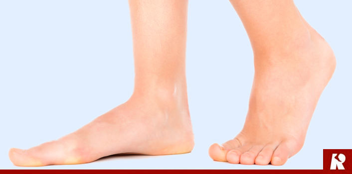
Causes
Women are affected by Adult Acquired Flatfoot four times more frequently than men. Adult Flatfoot generally occurs in middle to older age people. Most people who acquire the condition already have flat feet. One arch begins to flatten more, then pain and swelling develop on the inside of the ankle. This condition generally affects only one foot. It is unclear why women are affected more often than men. But factors that may increase your risk of Adult Flatfoot include diabetes, hypertension, and obesity.
Symptoms
Patients often experience pain and/or deformity at the ankle or hindfoot. When the posterior tibial tendon does not work properly, a number of changes can occur to the foot and ankle. In the earlier stages, symptoms often include pain and tenderness along the posterior tibial tendon behind the inside of the ankle. As the tendon progressively fails, deformity of the foot and ankle may occur. This deformity can include progressive flattening of the arch, shifting of the heel so that it no longer is aligned underneath the rest of the leg, rotation and deformity of the forefoot, tightening of the heel cord, development of arthritis, and deformity of the ankle joint. At certain stages of this disorder, pain may shift from the inside to the outside aspect of the ankle as the heel shifts outward and structures are pinched laterally.
Diagnosis
The history and physical examination are probably the most important tools the physician uses to diagnose this problem. The wear pattern on your shoes can offer some helpful clues. Muscle testing helps identify any areas of weakness or muscle impairment. This should be done in both the weight bearing and nonweight bearing positions. A very effective test is the single heel raise. You will be asked to stand on one foot and rise up on your toes. You should be able to lift your heel off the ground easily while keeping the calcaneus (heel bone) in the middle with slight inversion (turned inward). X-rays are often used to study the position, shape, and alignment of the bones in the feet and ankles. Magnetic resonance (MR) imaging is the imaging modality of choice for evaluating the posterior tibial tendon and spring ligament complex.
Non surgical Treatment
Treatment will vary depending on the degree of your symptoms. Generally, we would use a combination of rest, immobilization, orthotics, braces, and physical therapy to start. The goal is to keep swelling and inflammation under control and limit the stress on the tendon while it heals. Avoidance of activities that stress the tendon will be necessary. Once the tendon heals and you resume activity, physical therapy will further strengthen the injured tendon and help restore flexibility. Surgery may be necessary if the tendon is torn or does not respond to these conservative treatment methods. Your posterior tibial tendon is vital for normal walking. When it is injured in any way, you risk losing independence and mobility. Keep your foot health a top priority and address any pain or problems quickly. Even minor symptoms could progress into chronic problems, so don?t ignore your foot pain. 
Surgical Treatment
Good to excellent results for more than 80% of patients have been reported at five years' follow up for the surgical interventions recommended below. However, the postoperative recovery is a lengthy process, and most surgical procedures require patients to wear a plaster cast for two to three months. Although many patients report that their function is well improved by six months, in our experience a year is required to recover truly and gain full functional improvement after the surgery. Clearly, some patients are not candidates for such major reconstructive surgery.